


Spark® Cyto 多功能微量盤分析儀
Spark Cyto 是一款具有螢光成像和細胞檢測及全自動即時活細胞成像檢測系統的多功能微量盤分析儀,能夠即時控制實驗,使用並行資料獲取和分析,確保你離開實驗室時細胞不會停止活動,動態監控實驗從而不會錯失任何關鍵事件,為細胞學實驗提供深度有意義的見解。通過活體細胞成像與行業領先的檢測技術相結合,您能夠以前所未有的效率獲得獨特的多指標參數的定性和定量資料集合。 型錄下載
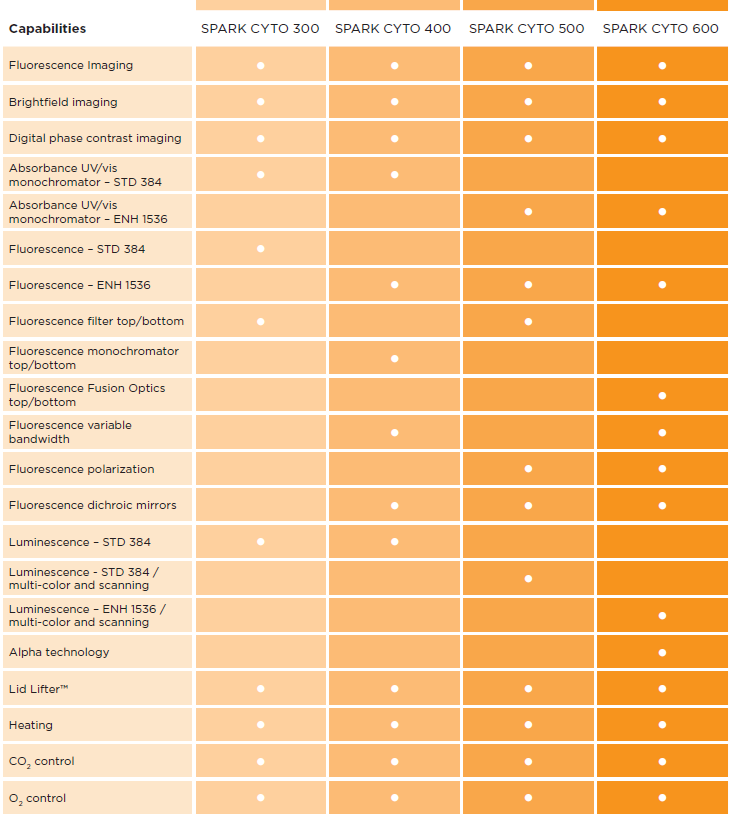
多種型號配置,滿足您的各種應用
Spark Cyto 有四種不同檢測方式的專有配置。
無論您選擇何種配置,他們都配備了可進行活細胞成像和檢測的全套系統和功能。
§即時檢測分析更多細胞§
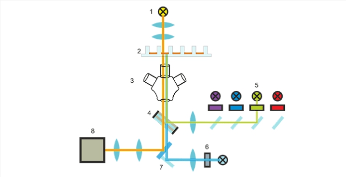
成像模組示意圖(1)明場LED光源;(2)樣本微孔板;(3)物鏡;(4)多帶通濾光片組;(5)螢光LED和激發濾光片組;(6)自動聚焦單元;(7)反光鏡;(8)CMOS相機
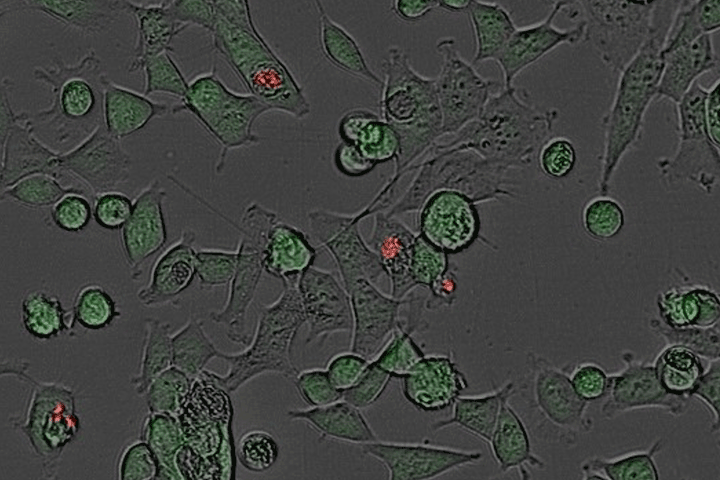
(1)專注於研究微孔板內活細胞的光學儀器
Spark Cyto 螢光成像模組設計在最少的使用者干預下為實驗提供清晰的圖像。
它使用三個不同的物鏡、五個LED(明場和螢光激發)、一個多波段濾光片組和
一個12位CMOS相機,Spark Cyto 消除了圖元偏移並快速提供高品質圖像。
- 一鍵選擇物鏡和通道
| Magnification | Numeric aperture | 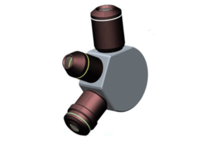 |
| 2x | 0.08 | |
| 4x | 0.13 | |
| 10x | 0.30 | |
| LED colors | Spectral range | |
| Blue | 381–450 nm | |
| Green | 461–530 nm | |
| Red | 543–611 nm | |
| Far red | 626–800 nm | |
| Bright field and digital phase contrast | ||
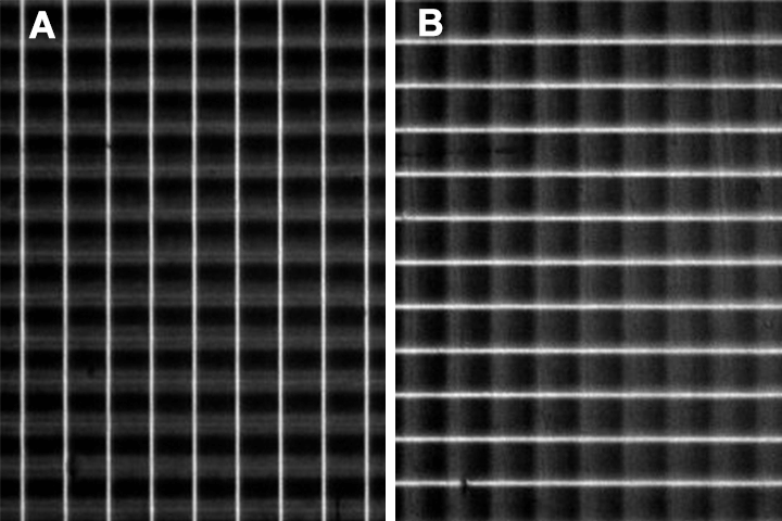
- 無像素位移的合成圖像
光學系統不需要移動光學元件或微孔板,就可以利用五個可用通道的任意組合完成微孔板成像。
這消除了像速移位,尤其是在使用兩個或多個通道時,顯著的提高圖像品質和獲取速度。
- 標配自動聚焦–專注於您的研究
自動對焦系統將一個擴展的網格圖案投射到樣品表面,這樣可以最大限度地減少孤立雜質
帶來的潛在扭曲。
- 現場觀看
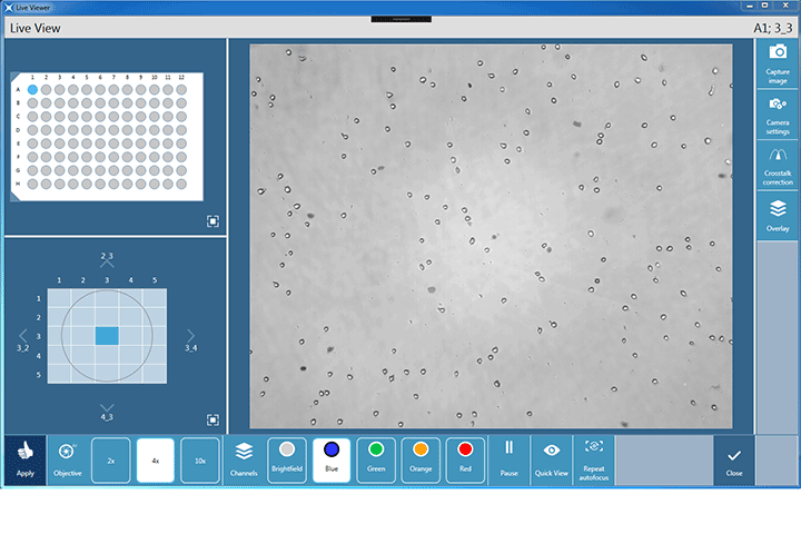
Live Viewer 軟體應用程式可以將 Spark Cyto 用作細胞圖像即時查看,即時監測。
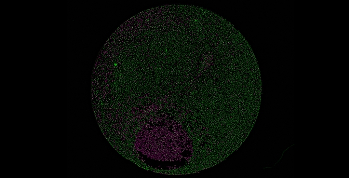
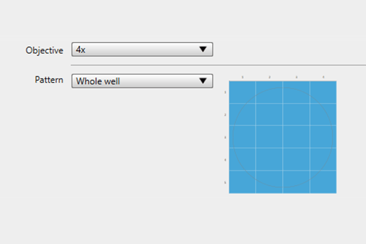
(2)96或384整孔成像分析
Spark-Cyto 彙集了一套專利申請中的獨特的寬視場細胞成像技術,以確保您可實現
真正的整孔細胞群研究。僅需一張圖像即可對96或384孔板進行整孔成像。沒有平鋪
或失真,這意味著讓你在更短的時間內全面瞭解細胞群,從而為您的研究帶來新方向。
- 一張圖片就能講述整個故事
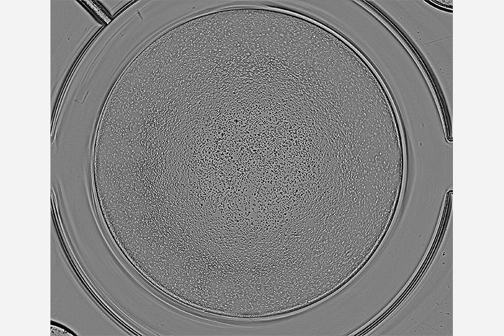
Spark-Cyto 用一張圖片捕捉整個微孔(96和384孔板格式),給你一張真實的研究圖片。
- 其他微孔板格式的整孔成像
spark cyto可在自動生成多個單一圖像的基礎上合成圖像,從而對這些應用進行整孔成像。
- 優化微孔板以獲得最佳結果
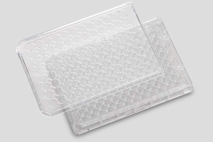
Spark Cyto 的成像功能經過專業優化,與TECAN的透明細胞培養板一起使用,從而
確保減少光學偽影並獲得最佳結果。
§測量更多參數§
Spark Cyto 的配置建立在Spark多功能微孔板檢測平臺之上,它將複雜的成像技術與
成熟的多模式微孔板檢測功能相結合,使您能夠定義新的研究方法,並比以往更快地
獲得可靠的資料。
§更好地控制實驗§
在不影響易用性和方便性的前提下,Spark cyto給您提供一個新的實驗控制水準,可用於
細胞檢測更廣泛的領域。五種常見細胞學應用的預定義方法提供了一種直觀的圖像獲取和
分析方法,並輔以“用戶定義”參數和即時實驗控制(Rec™)等附加功能,將為您實現解鎖
新的應用程式。
Spark Cyto 的功能 * 功能取決於Spark Cyto的配置
§典型應用§
- 核計數
- 轉染效率
- 細胞活力
- 細胞凋亡
- 匯合度評估
- 細胞遷移和傷口癒合
- ELISA實驗
- 微量DNA/RNA定量
- 核酸標記效率
- 蛋白定量
- 報告基因分析
- HTRF®, DELFIA®和LanthaScreen®
- Transcreener®
- DLR®
- BRET –包括NanoBRET®
§檢測模式§
- 螢光成像(藍色,綠色,紅色,遠紅色)
- 明場成像
- 數位相差成像
- 光吸收-包括紫外和可見光
- 螢光頂讀/底讀
- 時間分辨螢光(TRF)
- 各測量模式的全波長掃描功能
- FRET
- TR-FRET
- 螢光偏振(FP)
- 化學發光-輝光,閃光,多色,發光掃描
- AlphaScreen®, AlphaLISA®和AlphaPlex®
§其他性能§
- 帶攪拌和溫控的自動加樣器
- 濕度控制盒
- NanoQuant微量檢測板
- IQ/OQ服務的QC工具
- Spark-Stack微孔板堆疊
Typical performance values+
| Fluorescence imaging and cytometry | ||||
| Imaging technologies | Fluorescence, bright field, digital phase contrast | |||
| Imaging methods | Single color, multicolor, end-point, kinetics, whole-well | |||
| Sample formats | 6- to 384-well ANSI/SLAS-format microplates | |||
| Camera sensor | Grayscale, 5 Mpixel, CMOS Sony | |||
| Objectives | 2x (NA 0.08), 4x (NA 0.13), 10x (NA 0.30) | |||
| Optical properties | Objective | Pixel resolution | Optical resolution | Field of view |
| 2x | 3.45 μm | 4.50 μm | 8.47 x 7.09 mm | |
| 4x | 1.72 μm | 2.77 μm | 4.24 x 3.54 mm | |
| 10x | 0.69 μm | 1.20 μm | 1.69 x 1.42 mm | |
| Channels | Bright field, four fluorescence channels (blue, green, red, far-red) | |||
| Autofocus | Proprietary astigmatism-based technology | |||
| Field of view | Whole-well, 96- and 384-well imaging with a single image (2x and 4x objectives) | |||
| Applications | 4 pre-defined applications: Confluence, transfection efficiency, cell viability and cell death (apoptosis via annexin V-FITC), plus user-defined applications | |||
| Image collection rate | ≤12 min for 96-well plate, whole-well image with 2x, bright field and digital phase contrast ≤15 min for 96-well plate, center image with 10x, bright field, digital phase contrast + 1 fluorescence channel |
|||
| Analysis speed | ≤20 min for 96-well plate, whole-well image with 2x, bright field and digital phase contrast including real time confluence assessment | |||
| Fluorescence – enhanced | Fluorescence – standard | ||
| Light source | High energy xenon flash lamp | Light source | Dedicated xenon flash lamp |
| Spectral range | Ex: 230–900 nm
Em: 280–900 nm |
Spectral range | Ex: 230–900 nm
Em: 280–900 nm |
| Wavelength accuracy | Ex: <0.5 nm; Em: <0.5 nm | Wavelength accuracy | Ex: <1 nm; Em: <2 nm |
| Wavelength reproducibility | <0.5 nm | Wavelength reproducibility | <1 nm |
| Bandwidth | Adjustable from 5–50 nm | Bandwidth | Fixed @ 20 nm |
| Optical mirrors | 50 %, 510, 560, 625 nm built-in; 410, 430, 458, 593, 660 nm user-selectable dichroics | Optical mirrors | 50 %; 510 nm dichroic |
| Well scanning | Up to 100 x 100 data points | Well scanning | Up to 100 x 100 data points |
| FI (fluorescence intensity) Limit of detection1 | FI (fluorescence intensity) Limit of detection1 | ||
| Filter – top | ≤8 amol/well (10 μl; 1,536-well) | Filter – top | ≤25 amol/well (100 μl; 384 well) |
| Fusion* – top | ≤15 amol/well (10 μl; 1,536-well) | Fusion – top | ≤35 amol/well (100 μl; 384 well) |
| Mono – top | ≤20 amol/well (10 μl; 1,536-well) | Mono – top | ≤50 amol/well (100 μl; 384 well) |
| Filter – bottom | ≤180 amol/well (10 μl; 1,536-well) | Filter – bottom | ≤500 amol/well (200 μl; 96 well) |
| Fusion – bottom | ≤200 amol/well (10 μl; 1,536-well) | Fusion – bottom | ≤700 amol/well (200 μl; 96 well) |
| Mono – bottom | ≤220 amol/well (10 μl; 1,536-well) | Mono – bottom | ≤800 amol/well (200 μl; 96 well) |
| FP (fluorescence polarization)2 | FP (fluorescence polarization)2 | ||
| Spectral range | 300–850 nm | Spectral range | 300–850 nm |
| Precision – Filter | ≤1.25 mP | Precision – Filter | ≤1.5 mP |
| Precision – Fusion | ≤2.0 mP | Precision – Fusion | ≤2.5 mP |
| Precision – Mono | ≤2.5 mP | Precision – Mono | ≤3.0 mP |
| TRF (time-resolved fluorescence)3 | TRF (time-resolved fluorescence)3 | ||
| Limit of detection – Filter | ≤0.5 amol/well (20 μl; 384-well SV) | Limit of detection – Filter | ≤4.0 amol/well (100 μl; 384-well) |
| Limit of detection – Fusion | ≤0.6 amol/well (20 μl; 384-well SV) | Limit of detection – Fusion | ≤6.5 amol/well (100 μl; 384-well) |
| Limit of detection – Mono | ≤0.7 amol/well (20 μl; 384-well SV) | Limit of detection – Mono | ≤10 amol/well (100 μl; 384-well) |
| Fastest read time | Fastest read time | ||
| 384-well plate (FI) | ≤22 sec | 96-well plate (FI) | ≤13 sec |
| 1,536-well plate (FI) | ≤34 sec | 384-well plate (FI) | ≤30 sec |
| Absorbance (enhanced or standard) | |||
| Light source | Dedicated xenon flash lamp | ||
| Spectral range | 200–1,000 nm
OD range 0–4 OD |
||
| Scan speed (200–1,000 nm) | ≤5 sec | ||
| Wavelength accuracy | <0.3 nm | ||
| Wavelength reproducibility | ≤0.3 nm | ||
| Wavelength ratio accuracy (260/230) | <0.08 | ||
| Wavelength ratio accuracy (260/280) | <0.07 | ||
| Precision @ 260 nm | <0.2 % | ||
| Accuracy @ 260 nm | <0.5 % | ||
| Limit of detection (nucleic acids) | <1 ng/μl | ||
| Plate formats for all read modes – enhanced | Plate formats for all read modes – standard | ||
| 1-1,536 wells; NanoQuant Plate; Cuvettes; Roboflask® | 1-384 wells; NanoQuant Plate; Cuvettes; Roboflask | ||
| Luminescence (enhanced or standard) | |||
| Spectral range | 370–700 nm | ||
| Limit of detection – Glow4 | ≤225 amol/well (25 μl; 384-well SV) | ||
| Limit of detection – Flash5 | ≤12 amol/well (55 μl; 384-well) | ||
| Dynamic range | >9 orders of magnitude | ||
| Multi-color luminescence | 38 spectral filters; OD1, OD2, OD3 attenuation filters | ||
| AlphaScreen (enhanced or standard) | |||
| Limit of detection | <100 amol/well bio-LCK-P6; 20 μl
<2.5 ng/ml Omnibeads7; 20 μl |
||
| Uniformity | ≤3.0 % | ||
| Z´value | >0.9 | ||
| Fastest read times8 | ≤2 min (384-well plate)
≤1 min (96-well plate) |
||
| Gas Control Module (GCM™) | |||
| Adjustable concentration range – CO2 | 0.04–10 % (vol.) | ||
| Adjustable concentration range – O2 | 0.1–21 % (vol.) | ||
| Concentration accuracy – CO2 | <1 % (vol.) | ||
| Concentration accuracy – O2 | <0.5 % (vol.) | ||
| Reagent injectors | |||
| Syringe sizes | 0.5 ml; 1 ml | ||
| Pump speed | 100–300 μl/sec | ||
| Injection volume | 5–2,500 μl; step size: 1 μl | ||
| Dead volume | ≤100 μl | ||
| Injection accuracy and precision | ≤0.5 % at 450 μl | ||
| Temperature control | Ambient +3 °C up to 42 °C | ||
| Uniformity | <0.5 °C | ||
| Shaking | |||
| Linear, orbital, double-orbital; variable amplitudes and frequencies | |||
+Specifications are subject to change. Performance values represent the average observed factory tested values.
*Fusion Optics: a combination of filter and monochromator on the excitation and emission sides
1) Detection limit for fluorescein
2) FP detection limit @ 1 nM fluorescein
3) Detection limit for europium
4) Detection limit for ATP (144-041 ATP detection kit SL, BioThema)
5) Detection limit for ATP (ENLITEN® Kit)
6) (PE# 6760620; P-Tyr-100 assay kit)
7) (PE# 6760626D; Omnibeads)
8) Including temp. correction